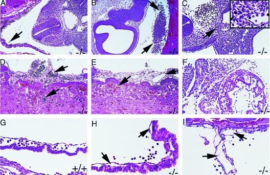
Figure 3.
Microscopic analysis of FII−/− embryos fixed and sectioned in situ within the uterine horns. (A) Section through a generally normal appearing E10.5 FII−/− embryo showing well-developed yolk sac vessels with blood filled lumens. However, there are occasional clusters of fetal blood cells (arrow) lying free in the yolk sac cavity. (B) Section from a FII−/− littermate of the embryo shown in A. This embryo shows a large pool of fetal blood cells in the yolk sac cavity (arrows). (C) Section from an E10.5 FII−/− embryo with extensive bleeding into the yolk sac cavity, but only small foci of tissue necrosis. (Inset) Small area of necrosis, indicated by arrow, at a higher magnification. (D) Section through the placenta from the embryo illustrated in A. The allantoic blood vessels and fetal vessels of the placenta are well developed and filled with fetal blood cells (arrows). (E) Section through the placenta from the embryo illustrated in B. Arrows indicate the allantoic and maternal sinusoids in the placenta. There are no fetal blood cells in the allantoic vessels and only maternal red blood cells are seen in the placenta. (F) Section from an E10.5 FII−/− embryo with widespread necrosis evident in the embryo. (G) Section showing the yolk sac membrane of an E10.5 FII+/+ embryo. Vessels are well developed, patent, and blood filled. (H) Section showing the yolk sac membrane from an E10.5 FII−/− embryo. Yolk sac vessels (arrows) are patent, but slightly collapsed and empty. Note fetal blood cells lying free in the yolk sac cavity. (I) Section through the vitelline vessel of an E10.5 FII−/− embryo that is well developed but empty. (A, B, D–F, 100×; C, G–I, 200×.)

