
Figure 1. 3-D and transverse images of the feline brain in vivo.
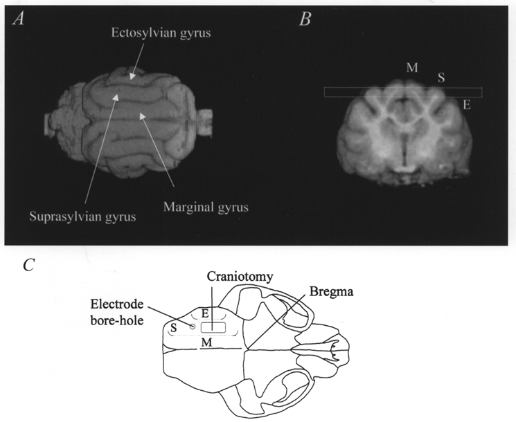
A shows a dorsal view of a volume-rendered reconstruction of the feline brain, in conjunction with a transverse image (B) showing the marginal (M), suprasylvian (S) and ectosylvian (E) gyri. The dashed box in B indicates the selection of the horizontal imaging slice (3 mm slice thickness) used for the DWEP and DWEPBOLD experiments described in the text. C reconstructs a dorsal view of the feline skull to indicate the surgical positions of the craniotomy and the electrode bore hole. Dotted lines indicate the relative positions of the three gyri.