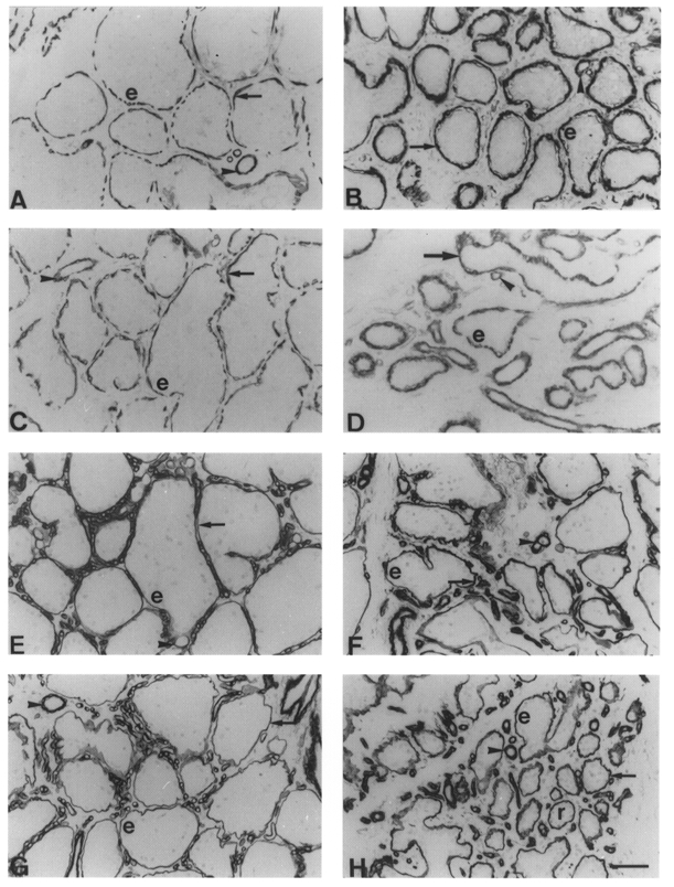
Figure 6. Immunocytochemical staining of myosin and actin in goat mammary glands milked once and thrice daily.

A-D, smooth muscle myosin staining with MAb hSV-Min in glands milked thrice daily (A and C) or once daily (B and D) for 4 weeks (A and B) or 10 weeks (C and D). Arrows, myoepithelial cells; arrowheads, blood vessels; e, epithelial cells. E-H, type IV collagen staining with MAb CIV22 in glands milked thrice daily (E and G) or once daily (F and H) for 4 weeks (E and F) or 10 weeks (G and H). Arrows, basement membrane; arrowheads, blood vessels; e, epithelial cells; r, resting ductules. Scale bar, 50 μm; applies to all panels.
