
Figure 3. Effects of K+ channel antagonists on Purkinje cell IPSCs.
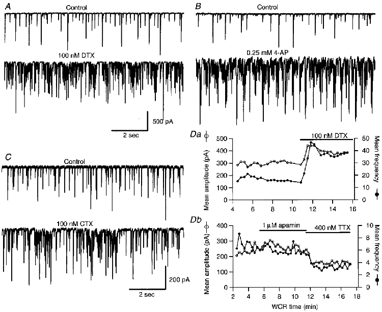
A-C, representative traces of spontaneous IPSCs recorded from three Purkinje cells before (upper panels) and after (lower panels) the addition of 100 nM αDTX (A), 0.25 mm 4-AP (B) and 100 nM CTX (C). The calibration bars in A apply also to the experiment shown in B. D, plots of the mean IPSCc amplitude (○) and mean frequency (•) calculated for 20 s time bins, as a function of whole-cell recording (WCR) time. Da, shows an experiment in which αDTX was applied, while in the experiment shown in Db apamin was tested. NBQX (10 μm), APV (100 μm) and bicuculline (1 μm) were present throughout the recordings shown in A-C as well as in the experiment shown in Da. In the experiment shown in Db 10 μm NBQX and 100 μm APV were present, but no bicuculline was added to avoid possible effect of this drug on apamin-sensitive K+ channels (Seutin et al. 1997).