
Figure 1. Schematic drawing of the experimental set-up.
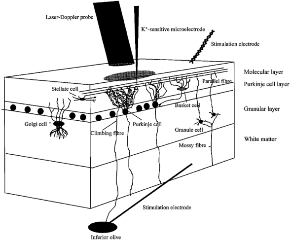
A three-dimensional sagittal view of the rat cerebellar cortex, showing the neurones of interest and the position of the laser-Doppler probe, K+-sensitive microelectrode and stimulating electrodes. The superficial parallel fibres were stimulated by a bipolar electrode positioned on the cerebellar surface, while the climbing fibres were stimulated by a monopolar electrode lowered into the inferior olive. CeBF was recorded by a laser-Doppler flowmetry (LDF) probe located 0.5 mm above the pial surface, whereas changes in [K+]o were recorded with a K+-sensitive microelectrode lowered 50-100 μm into the cortex.