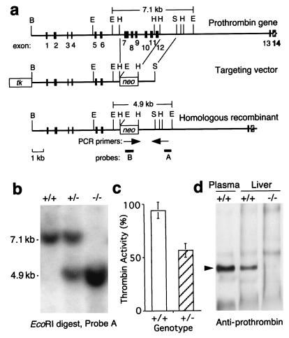
Figure 1.
Targeting of the prothrombin gene by homologous recombination. (a) Structure of mouse prothrombin gene and targeting vector. The cloned genomic DNA fragment extended from the first BamHI site to the last shown EcoRI site. Solid rectangles represent exons 1–12. Additional exons 13 and 14 (hatched rectangles) are indicated based on the structure of the human prothrombin gene. Cleavage sites are shown for BamHI (B), EcoRI (E), HindIII (H), and SalI (S). The targeting vector contains a pgk-neo cassette in place of exons 7–12 and a pgk-tk cassette. The product of homologous recombination is shown at the bottom. Positions of PCR primers (arrows) and hybridization probes (A and B) used to detect successful gene targeting are indicated. The EcoRI digest will generate a 7.1 kb fragment from the untargeted allele and a 4.9 kb fragment from the targeted allele, both of which are recognized by probe A. (b) Southern blot of EcoRI-digested tail DNA from Cf2+/+, Cf2+/−, and Cf2−/− littermates hybridized with probe A. (c) Prothrombin levels in mouse plasma sampled from 10 mice of each genotype (mean ± SD). (d) Western blot of prothrombin in mouse liver extracts. Arrowhead indicates the position of 72 kDa prothrombin.

