
Figure 2.
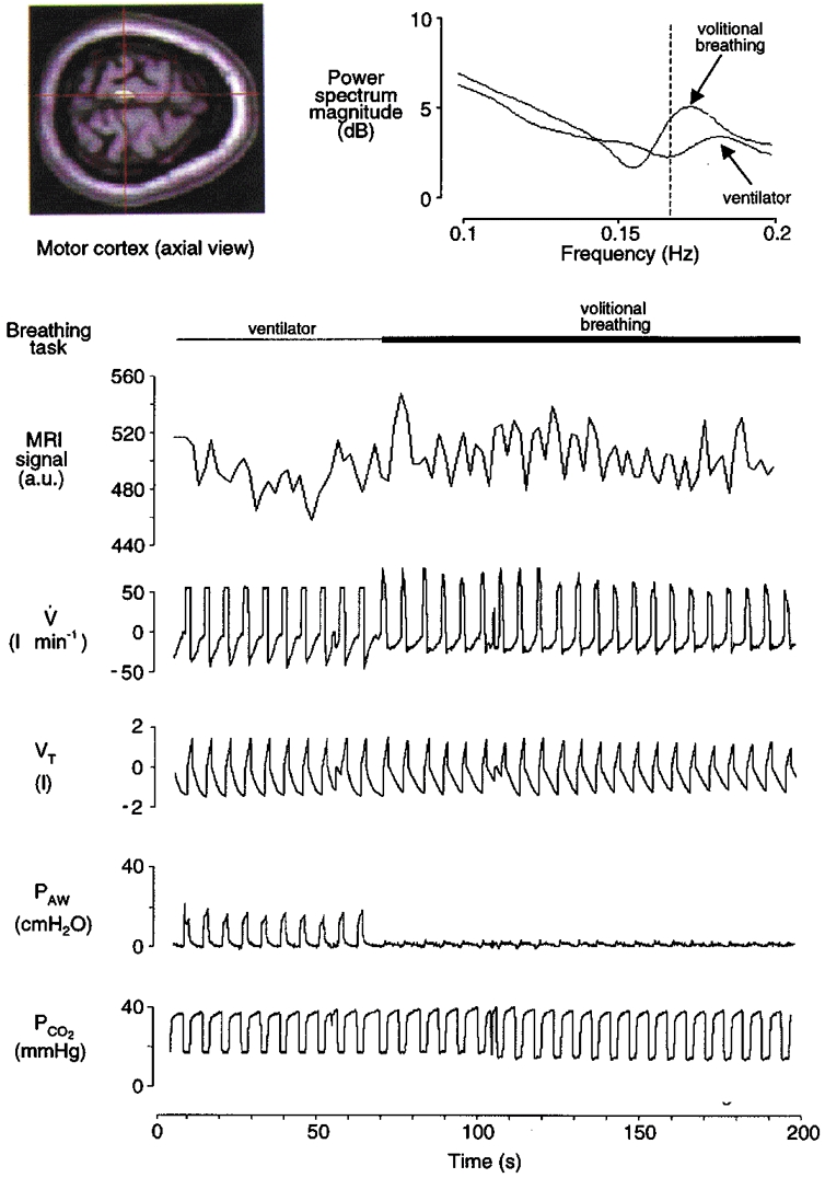
Record from a typical subject (F) during mechanical ventilation (passive task; first minute only) and volitional inspiration (active task; remainder of scan series). The left inset depicts an axial fMRI image and a region of interest in the left motor cortex (centre of cross-hairs). The fMRI signal (in arbitrary units, a.u.) at this region is time-aligned with the respiratory waveforms: V, airflow; VT, tidal volume; PAW, airway pressure; PCO2, partial pressure of end-tidal CO2. (NB, the ‘raw’ fMRI signal in the figure is unadjusted for haemodynamic lag.) There was a small average increase in fMRI signal during volitional inspiration relative to passive ventilation in this superior region of motor cortex. There were also phasic fMRI changes synchronised with inspiration and expiration that are particularly evident during the active task. This synchronisation (between neural activation and volitional inspiration) is depicted in the power spectra of these fMRI signals (right inset). The fMRI spectrum during volitional inspiration has an isolated peak near the frequency of breathing (0.167 Hz; vertical dashed line), whereas the spectrum during passive mechanical ventilation shows less specificity with breathing frequency.