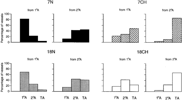
Figure 5. Categories of vessels that branched from primary arterioles and secondary arterioles in 7N, 18N, 7CH and 18CH rats.

Each set of histograms shows the percentage of branches from primary arterioles (1°A, lefthand set) or secondary arterioles (2°A, righthand set) that were identified as 1°A, 2°A and terminal arterioles (TA) on the basis of internal diameter. The total number of branches considered was 60–80 for both primary and secondary arterioles in the 6 spinotrapezius muscles of each group of rats. Shading of columns for each group of rats is as in Fig. 3.
