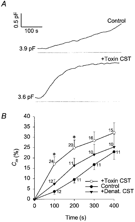
Figure 2. Time-dependent changes in membrane capacitance with high [Ca2+]i.

A, changes in membrane capacitance in rat melanotrophs elicited with cytosol dialysis with 1 μm . The top trace is the control and the trace below is from a CST-treated cell (15 nm Sa and 15 nm Sb, treated for 1 h). Values adjacent to traces indicate resting capacitance. B, mean changes in membrane capacitance (%Cm) relative to resting Cm with high [Ca2+]i. Changes in membrane capacitance are determined every 100 s after the start of cytosol dialysis with 1 μm . *Significant differences between the control and CST-toxin treated cells (Student's t test, P < 0.01). ○, CST toxin treated cells (+Toxin CST); ▾, cells treated with the temperature-denatured CST toxin (15 nm Sa and 15 nm Sb, treated for 1 h (+Denat. CST); •, control cells. Numbers adjacent to points indicate numbers of cells tested, bars indicate s.e.m.
