
Figure 1. Effect of catalase in the presence of NO donors in frog cardiac fibres.
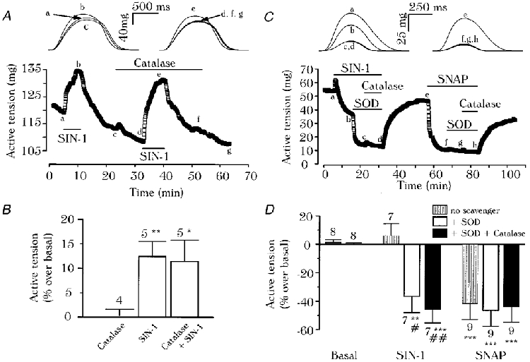
A ventricular (A) or an atrial (C) fibre was initially superfused with control solution. In A, SIN-1 (100 μm) was applied in the absence or presence of catalase (1000 U ml−1), as indicated by the lines. In C, SOD (50 U ml−1) and catalase (200 U ml−1) were added in the presence of SIN-1 (100 μm) or SNAP (100 μm). Top, traces were recorded at the times indicated by the corresponding letters on the main graphs. B, summary of the effects of catalase (1000 U ml−1) and SIN-1 (100 μm), alone or in combination, in ventricular fibres where SIN-1 had induced positive effects. D, summary of the effects of SNAP (100 μm) or SIN-1 (100 μm) in the presence of SOD (50-200 U ml−1), with or without catalase (200-1000 U ml−1). In B and D, the active tension in the presence of agents was normalised to its value in the absence of agents. Bars and lines are the mean ±s.e.m. of the number of experiments indicated. Statistical differences from the basal level (*) or from SIN-1 (#) are indicated as *#P < 0.05; **##P < 0.01; ***P < 0.005.