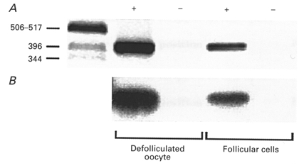
Figure 6. RT-PCR amplification of XlmR transcripts in follicular cells.

A, ethidium bromide-stained gel of RT-PCR-amplified products. The left-hand lane contains DNA markers (in bp). +, cDNA from defolliculated oocytes or isolated follicular cells. -, negative controls, RNA without reverse transcriptase. B, Southern blot of the same gel as in A, hybridized with XlmR probe.
