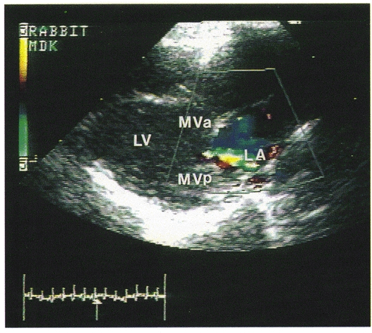
Figure 1. An example of a colour Doppler echocardiograph in a rabbit with mitral regurgitation.

The picture is a frozen frame during ventricular systole (as indicated by the arrow pointing to the ECG tracing at the bottom left) when the blood is regurgitating back to the left atrium, which is seen as a blue jet inside the left atrium. LV, left ventricle; LA, left atrium; MVa, mitral valve (anterior cusp); MVp, mitral valve (posterior cusp).
