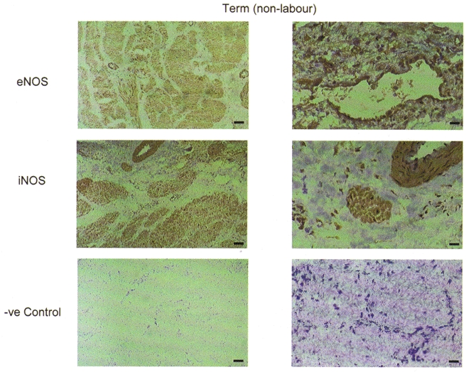
Figure 4. Immunohistochemical staining of sections of human pregnant myometrial tissue with monoclonal anti-NOS antibodies.

Representative sections are shown of term non-labouring tissue. Cryostat sections 5 μm thick were incubated with monoclonal antibodies raised against eNOS and iNOS. Negative controls were incubated with non-immune mouse IgG. Staining was by avidin-biotin-peroxidase.
