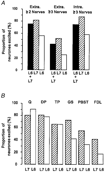
Figure 7. Convergence from group II muscle afferents onto individual interneurones.

A, histograms showing the proportion of neurones discharged by group II afferents of two or more muscle nerves (left) and three or more muscle nerves (middle), and the proportion of neurones in which EPSPs were evoked by group II afferents of three or more muscle nerves (right). The filled bars represent neurones in both the L6 and L7 segments, the hatched bars neurones in the L7 segment and the open bars neurones in the L6 segment. Note the greater convergence in L7 than in L6. B, histograms showing a comparison of the proportions of interneurones in the L7 and L6 segments in which EPSPs were evoked by group II afferents of different nerves. The hatched bars represent neurones recorded in the L7 segment and the open bars represent neurones recorded in the L6 segment. Note that group II afferents of PBST, GS and FDL excited a larger proportion of neurones in the L7 segment than in L6, while afferents of Q, DP and TP were similarly effective in the two segments.
