
Figure 1. Enhanced myogenesis by implanted myoblasts in irradiated muscle.
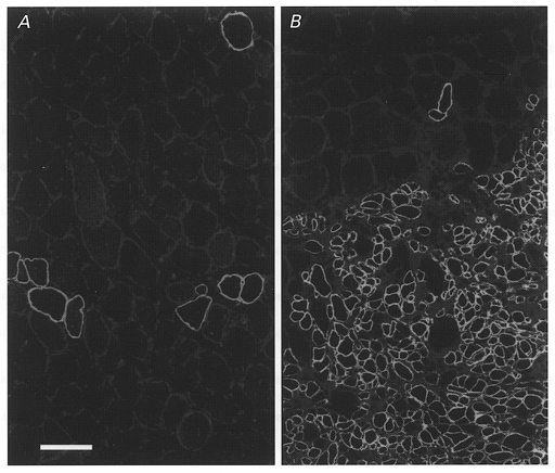
Dystrophin expression in representative transverse sections from the non-irradiated left soleus (A) and the irradiated right soleus (B) of a dystrophin-deficient mdx mouse 67 days after transplantation of myoblasts derived from a non-dystrophin deficient C57Bl/10 mouse. No muscle cryodamage had been imposed before implantation. A, a low number of large- and small-diameter muscle fibres in the non-irradiated muscle express dystrophin. B, dystrophin expression in numerous small-diameter fibres of donor origin is seen in the irradiated soleus muscle. Scale bar in A represents 50 μm and applies also to B.