
Figure 3. Donor myoblasts re-build irradiated/damaged host muscles.
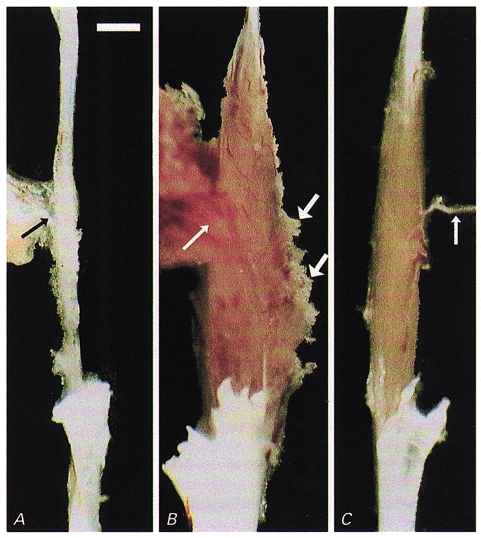
Macroscopic appearance of soleus muscles dissected with nerves (thin arrows) for force recordings. A, a fibrotic remnant (5 mg) of a Balb/c soleus muscle irradiated and cryodamaged 96 days previously which produced no force upon supramaximal stimulation and contained no muscle fibres. B, a large muscle (21 mg) had grown in the bed of an irradiated, damaged and cell-implanted muscle of another animal within 97 days after implantation. The muscle produced 45 mN maximum force upon electrical stimulation. Muscle tissue extruding from the muscle belly (thick arrows) had been damaged during dissection. C, for comparison, the untreated soleus contralateral to that in B is shown (10 mg, 177 mN). Scale bar in A represents 2 mm and applies also to B and C.