
Figure 4. Histological appearance of a muscle repopulated by donor cells.
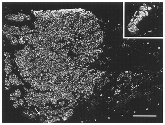
Immunofluorescent staining of desmin in a transverse section through the mid-belly of an irradiated/damaged and cell-implanted muscle studied 3 months after treatment. Contiguous and aligned small-diameter muscle fibres occupy about one-half of the muscle cross-sectional area (left half of the large panel). Fibres of large diameter (one at arrow) are very rare. The rest of the area (muscle border marked by asterisks) contains loose fibrous and fatty tissue (desmin negative) and very few desmin-positive muscle fibres. Inset, desmin-stained transverse section through the mid-portion of an irradiated/ damaged non-implanted muscle studied 4 months after treatment. Only a few muscle fibres, mostly of large calibre, are present in the soleus muscle remnant. Calibration bar represents 200 μm and applies also to the inset.