
Figure 5. Y chromosome-positive nuclear progeny of the implanted cells.
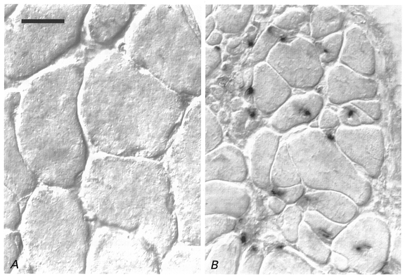
In situ hybridisation of a Y chromosome-specific cDNA probe on transverse sections of an irradiated/damaged muscle implanted with myoblasts derived from a male donor (B) and the contralateral intact soleus (A) of a female Balb/c mouse 4 months after treatment. No nuclear staining is present in or between muscle fibres of normal diameters in the intact muscle of the female animal (A). B, numerous donor-derived positive nuclei are incorporated into muscle fibres most of which have small diameters. Pictures were taken using differential interference contrast (Nomarsky) optics. Scale bar in A represents 20 μm and applies also to B.