
Figure 1. Alignment of various transmitter-gated ion channel M2 regions.
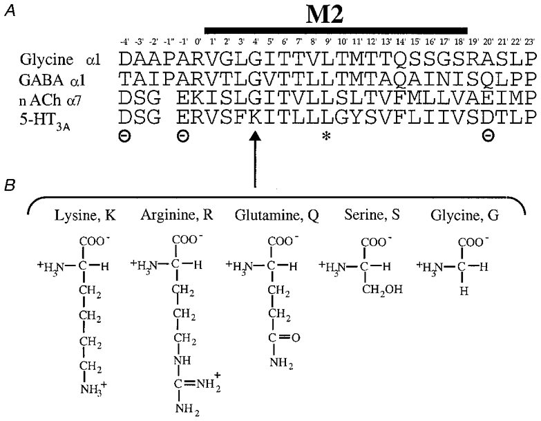
A, the amino acid sequence (single letter code) of the M2 region of the murine 5-HT3A receptor (Maricq et al. 1991) is shown aligned with the corresponding regions of the glycine (α1, Grenningloh et al. 1987) GABAA (α1, Schofield et al. 1987) and nACh (α7, Couturier et al. 1990) receptors. The asterisk indicates the position of the conserved leucine (9′L) residue involved in channel gating (Filatov & White, 1995; Labarca et al. 1995). The positions of the cytoplasmic (–4′), intermediate (–1′) and extracellular (20′) rings of charged residues bordering M2 in the cationic channels are also indicated. B, the amino acids substituted for 4′lysine (4′K, arrow) in this study: arginine (R), glutamine (Q), serine (S) and glycine (G) shown to illustrate the differences in their side chain charge and length.