
Figure 4. Effect of extracellular Ca2+ on desensitization of the 5-HT3A receptor.
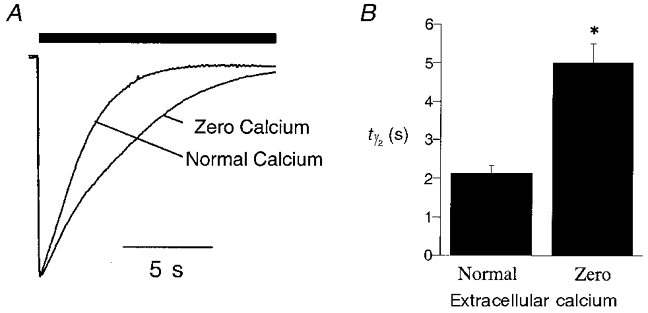
The rate of desensitization of WT receptor whole cell currents was studied in ‘normal’ (E1; 1.8 mM calcium) or ‘zero’ calcium (∼10 nM) extracellular solutions (see Methods). A, sample traces showing the effect of extracellular Ca2+ on the rate of desensitization of WT receptor whole cell currents in the continued presence of a maximal concentration of agonist (30 μm 5-HT, filled bar). B, graph illustrating the difference in the rate of desensitization of whole cell current recorded in ‘normal’ or ‘zero’ extracellular Ca2+. The data represent the time taken for the current to decay to half of its peak value in the continued presence of agonist (i.e. half-time of desensitization, t1/2; n = 10–12). *Statistically different from responses evoked in normal extracellular solution (P < 0.05).