
Figure 7. Time course of depolarization- and caffeine-induced change in rhod-2 fluorescence.
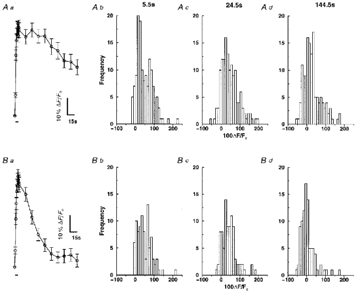
Smooth muscle cells were loaded with 1–1.5 μM rhod-2 AM for 1 h to enable monitoring of [Ca2+]m. Aa, time course of the change in mitochondrial rhod-2 fluorescence during a brief train of depolarizations. Approximately 20–30 mitochondria were selected in each cell and the brightest pixel was then followed through time (n= 7 cells, 159 mitochondria). The bar beneath the graph represents the period of depolarizing stimulation. Ab-d, histograms of the change in rhod-2 fluorescence at t= 5.5, 24.5 and 144.5 s for the depolarizing stimulus. Ba, time course of the change in mitochondrial rhod-2 fluorescence during a 5 s caffeine application. Approximately 20–30 mitochondria were selected in each cell and the brightest pixel was then followed through time (n= 5 cells, 115 mitochondria). The bar beneath the graph represents the period of caffeine stimulation. Bb-d, histograms of the change in rhod-2 fluorescence at t= 5.5, 24.5 and 144.5 s for stimulation with caffeine.