
Figure 4. Western blots of homogenate, plasma membrane and vesicles of Xenopus oocytes.
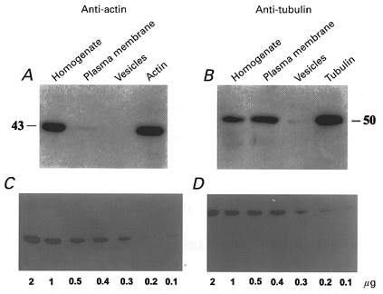
Actin (A) and tubulin (B) were labelled with anti-actin and anti-tubulin antibodies, respectively. The resolution limits of the technique were calibrated with different concentrations of actin (C) and tubulin (D).