
Figure 5. Transmission and scanning electron microscopy of oocyte surface and plasma membrane vesicles.
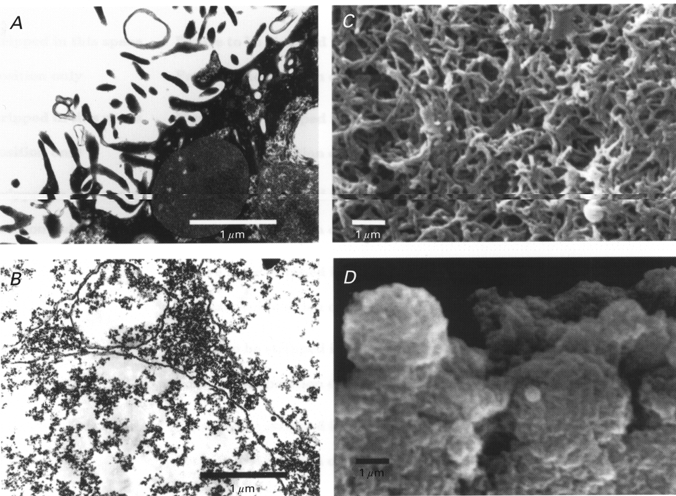
A, TEM of a portion of the oocyte surface showing prominent microvilli containing dark cytoplasmic material. B, TEM of PMVs indicating a smooth, even trilaminar membrane, with no evidence of particles or fibres associated with the cytoplasmic side of the membrane. C, SEM of the oocyte surface indicating the high density of microvilli. D, SEM of PMVs indicating a basically smooth surface membrane with no appendages.