
Figure 1. Northern blot analysis of MyHCneo mRNA.
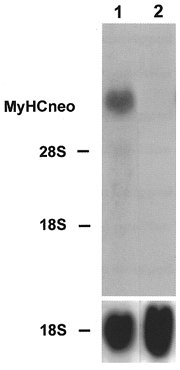
The rabbit primary skeletal muscle culture cells were grown for 11 (lane 1) and 22 days (lane 2). Total RNA (20 μg) was isolated at the time points indicated, fractionated on a 1.2 % agarose-formaldehyde gel, and transferred to nitrocellulose. The blots were hybridized with the 32P-labelled 3′ terminal PstI fragment of perinatal MyHC cDNA (1 × 10⁶ c.p.m. ml−1) or an rDNA probe from 18S rRNA (1 × 10⁶ c.p.m. ml−1). The positions of 18S rRNA (1.9 kb) and 28S rRNA (4.8 kb) on the ethidium bromide-stained gel are indicated.