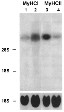
Figure 4. Effect of early addition of Ca2+ ionophore on the expression of slow MyHCI mRNA and fast MyHCII mRNA.

Cultures were grown for 24 days without ionophore (lanes 1 and 3) or from day 11 for a further 13 days with Ca2+ ionophore A23187 (4 × 10⁻⁷ M) (lanes 2 and 4). Total RNA (20 μg) was isolated from control and ionophore treated cultures at the time points indicated, fractionated on a 1.2 % agarose-formaldehyde gel, and transferred to nitrocellulose. Lanes 1 and 2 were probed with the 32P-labelled 3′ terminal HinfI fragment of MyHCI cDNA (1 × 10⁶ c.p.m. ml−1) or an 18S rDNA probe (1 × 10⁶ c.p.m. ml−1). Lanes 3 and 4 were probed with the 32P-labelled 3′ terminal PstI fragment of MyHCIId cDNA (1 × 10⁶ c.p.m. ml−1) or an rDNA probe from 18S rRNA (1 × 10⁶ c.p.m. ml−1). The positions of 18S rRNA (1.9 kb) and 28S rRNA (4.8 kb) on the ethidium bromide-stained gel are indicated.
