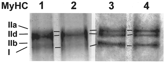
Figure 5. Electrophoresis of MyHC isoforms.

Electrophoresis of myosin extracts from myotubes growing on microcarriers. MyHC of the control on day 24 (lane 1) and day 29 (lane 2) and isoform pattern of the Ca2+ ionophore A23187 (4 × 10⁻⁷ M) treated cells after 2 (lane 3) and 7 days (lane 4) of incubation.
