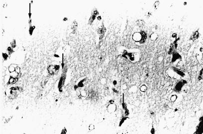
Figure 6. Photomicrograph of the anterior portion of the cingulate cortex in the right cerebral frontal lobe of dog 6. Notice acute nerve cell change (arrows), swelling of astrocytic nuclei, and mild vascular endothelial swelling. Acute nerve cell change consists of a shrinkage of nerve cell bodies with eosinophilic and homogeneous cytoplasm and atrophic nuclei. Hematoxylin and eosin stain; magnification: 340X.

An official website of the United States government
Here's how you know
Official websites use .gov
A
.gov website belongs to an official
government organization in the United States.
Secure .gov websites use HTTPS
A lock (
) or https:// means you've safely
connected to the .gov website. Share sensitive
information only on official, secure websites.
