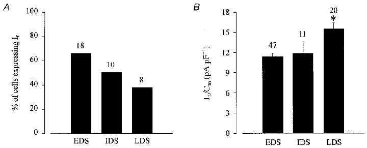
Figure 2. Functional expression of If in ES cell-derived cardiomyocytes at different stages of development.

A, the percentage of cells expressing If declined during cardiomyogenesis. B, at later stages of development an increase in the current density (If/Cm, where Cm is membrane capacitance) of If was observed. If was evoked by applying a 2 s hyperpolarizing voltage step from a holding potential of −35 to −110 mV (*statistically significantly different to EDS and IDS at the P < 0.05 level using Student's unpaired t test). Numbers above bars are numbers of replicates.
