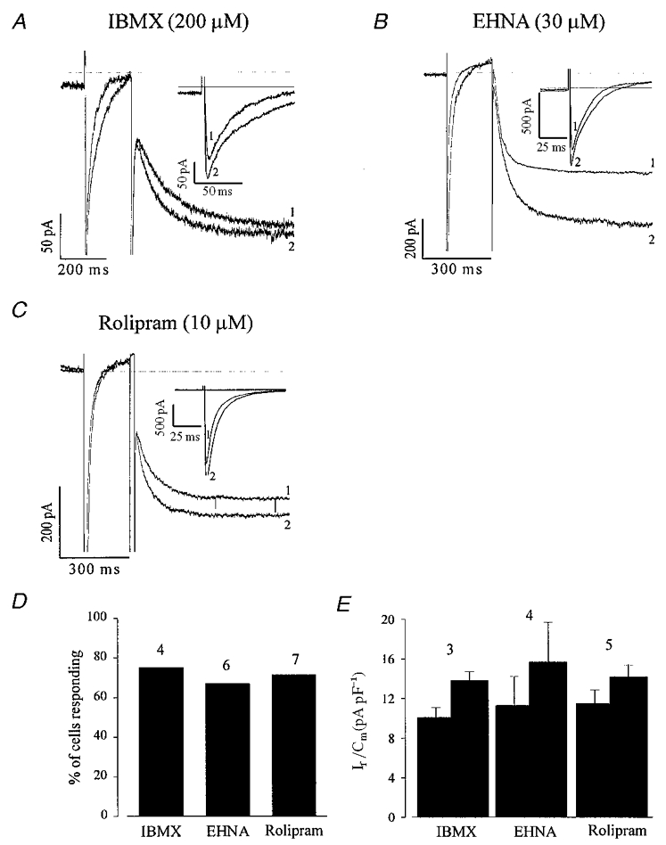
Figure 7. The non-selective phosphodiesterase (PDE) inhibitor IBMX as well as PDE2 and PDE4 antagonists stimulate If and ICa,L in EDS cardiomyocytes.

A–C, the non-selective antagonist IBMX (200 μM; A), the PDE2 selective antagonist EHNA (30 μM; B) and the PDE4 antagonist rolipram (10 μM; C) stimulate both If and ICa,L in EDS cardiomyocytes. The same voltage protocol as in Fig. 6 was used. The insets display ICa,L at a more extended time scale. Due to the slight delay in the maximal response of If as compared with ICa,L and the fast run down of the latter, the ICa,L traces displayed in B and C were recorded prior to If. D shows that most of the EDS cardiomyocytes tested responded to PDE antagonist application with a stimulation of If and ICa,L. E shows that an increase in If density was also observed under these conditions.
