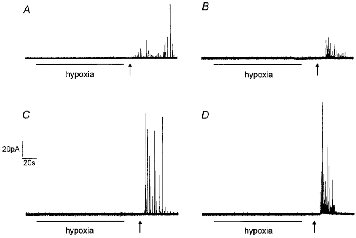
Figure 1. Hypoxia does not evoke catecholamine release from isolated rat type I carotid body cells.

Example amperometric recordings from type I cells exposed to hypoxia (PO2 8–14 mmHg) for the period indicated by the horizontal bar, and then to normoxic solution containing 50 mM K+, beginning at the time point indicated by the arrow. Cells were either perfused at 21–24 °C (A and C) or 35–37 °C (B and D) with extracellular solution buffered either with Hepes (A and B) or HCO3−/CO2 (C and D). Note the lack of secretory response to hypoxia in all cases. The presence of an intact secretory apparatus was confirmed in each cell by the application of 50 mM K+. Scale bars apply to all traces.
