
Figure 1. Rat sinoatrial (SA) node tissue after enzyme treatment (A) and a myocyte isolated by enzyme treatment (B).
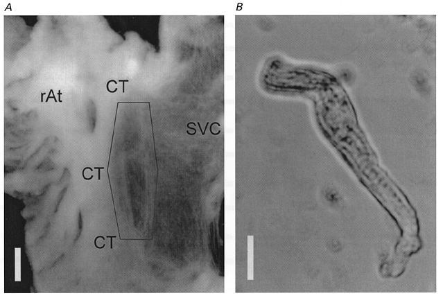
A, atrial tissue after enzyme treatment was mounted on the bottom of a chamber with the endocardial side up. The crista terminalis (CT), right atrium (rAt) and thin vein wall of superior vena cava (SVC) were clearly visible even after the enzyme treatment. The hexagon indicates the area from where the tissue was dissected to dissociate the SA node cells. The calibration bar is 1 mm. B, the SA node cell in the recording chamber was spindled shaped and showed spontaneous action potentials with slow diastolic depolarization. The calibration bar is 10 μm. The cell ends are out of focus because both cell ends bent upward from the bottom of the recording chamber.