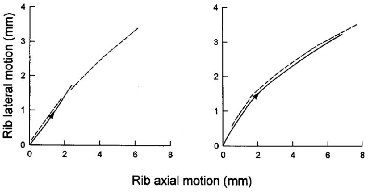
Figure 2. Pattern of rib displacement during inspiration.

Records obtained in a representative animal in the control condition (left panel) and after section of the phrenic nerves in the neck (right panel). The continuous line in each panel corresponds to spontaneous inspiration, and the dashed line corresponds to mechanical ventilation. Note that the pattern of rib motion during spontaneous inspiration was very close to that seen during mechanical ventilation both before and after phrenic nerve section.
