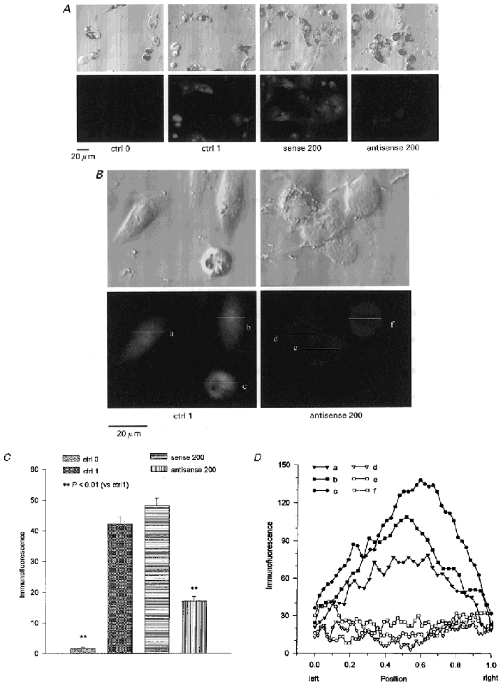
Figure 7. ClC-3 immunofluorescence.

The ciliary epithelial cells were incubated in control solution (ctrl; no additives), 200 μg ml−1 ClC-3 sense (sense 200) or 200 μg ml−1 ClC-3 antisense (antisense 200) oligonucleotides with the transfecting agent lipofectin (20 μg ml−1) for 48 h. The ClC-3 immunofluorescence in the absence (ctrl 0) or presence (ctrl 1, sense 200, antisense 200) of anti-ClC-3 antibody, is represented as a grey level (8-bit scale, 0 = black, 255 = white). The images in the upper row are transmitted light micrographs of the same images as the laser scanning confocal immunofluorescence images in the lower row (in both A and B). The ClC-3 immunofluorescence in the NPCE cells was reduced by the antisense (A, far right; B, right) but not by the sense oligonucleotides. The majority of ClC-3 immunofluorescence was inside the cells (typically in the nuclear area) and it was significantly diminished by the ClC-3 antisense treatments (B). C, the ClC-3 immunofluorescence of NPCE cells under different treatments expressed in units of grey level. D, the immunofluorescence distribution along the lines (a, b, c, d, e, f) indicated in B. The X-axis shows the scanning position of the lines. Position 0 is the left end of the lines and 1 is the right end.
