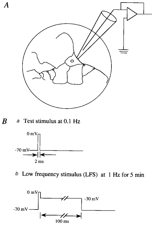
Figure 1. Schema showing preparations of a solitary neurone to which a whole-cell patch electrode was attached (A) and stimulus procedures (B).

A, schematic depiction of glial microisland (circle) and a solitary neurone grown on the island. Autapses are schematically indicated by small circles. Ba, test stimuli were given to the soma every 10 s. Bb, conditioning stimuli were paired with the depolarisation to −30 mV for 100 ms and repeated at 1 Hz for 5 min.
