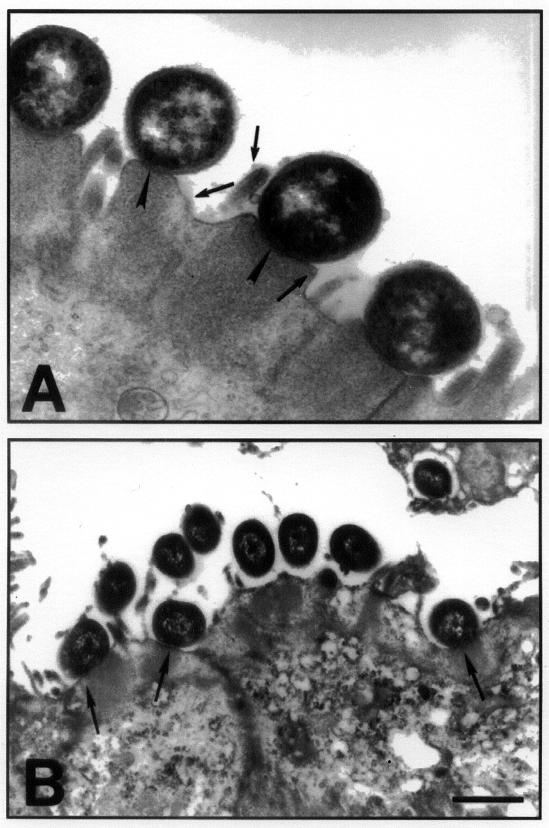
Figure 4. Transmission electron micrograph of an eae-positive and eae-negative STEC associating with the colonic epithelium. A. The figure shows the classical AE lesion in an area of the colon infected with STEC O5:NM. Note the pedestal formation and the close association of bacteria with the colonic epithelial cells (arrowheads) and the effacement of microvilli (arrows in the lumen). B. Transmission electron micrograph of colonic mucosa infected with an eae-negative STEC of serotype O113:H21 shows that some bacteria are surrounded by an electronlucent zone and are at some distance from the epithelium. However, other bacteria (arrows) appear to be associated with typical AE lesions.

An official website of the United States government
Here's how you know
Official websites use .gov
A
.gov website belongs to an official
government organization in the United States.
Secure .gov websites use HTTPS
A lock (
) or https:// means you've safely
connected to the .gov website. Share sensitive
information only on official, secure websites.
