
Figure 2. Reconstruction of a somatostatin-immunopositive O-LM to pyramidal cell pair.
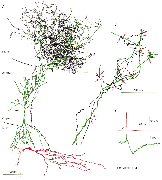
A, extensive overlap between the distal dendrites of the pyramidal cell (green) and the interneurone axon (black) at P15. The soma and dendrites of the interneurone are shown in red. B, light microscopic analysis revealed seventeen axon to dendrite close appositions (red arrows). C the somatically recorded postsynaptic current in the pyramidal cell was very small (cf. Fig. 1).