
Figure 4. Reconstruction and synaptic effect of a somatostatin-immunopositive O-BiC on a pyramidal cell at P15 (see also Fig. 5).
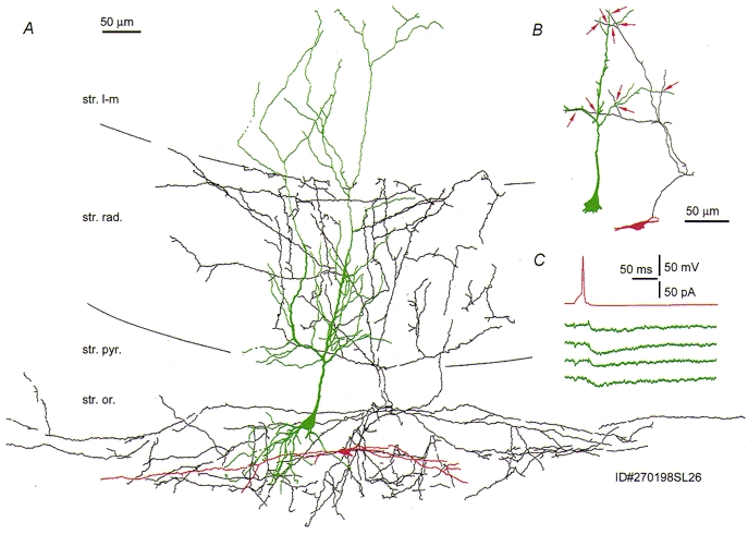
A, the axon (black) of the interneurone (soma and dendrites in red) is largely restricted to str. oriens and radiatum and overlaps the dendritic tree of the postsynaptic pyramidal cell (green). Note the sparing by the axon of str. pyramidale, and the dendritic tree restricted to str. oriens. B, close appositions (red arrows) of the interneurone axon and the pyramidal dendritic tree. All but one of the appositions are provided by one main axon collateral. C, single action potentials in the interneurone evoked slow uIPSCs (single sweeps) in the postsynaptic pyramidal cell.