
Figure 8. Perisomatic innervation of pyramidal cells by axo-axonic and basket cells.
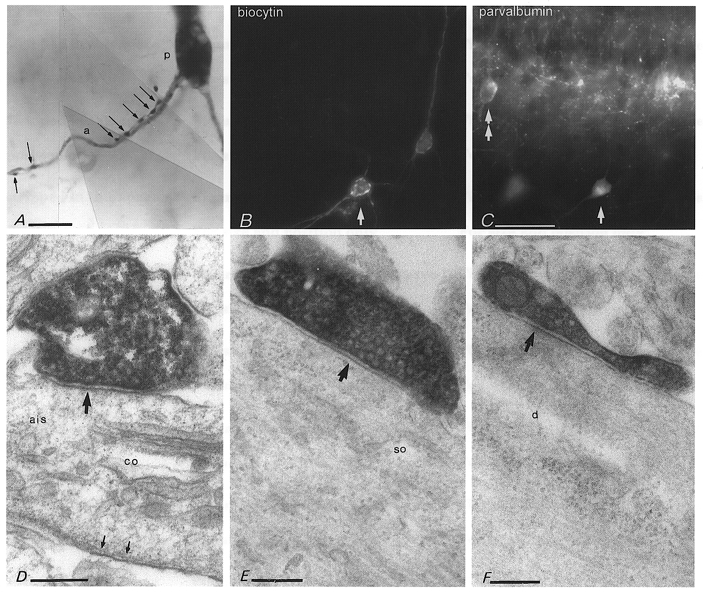
A, an axo-axonic cell (P13) innervates the axon initial segment (a) of a pyramidal cell (p) at two distinct regions via two axon collaterals providing at least seven boutons (arrows). B and C, parvalbumin immunoreactivity in another axo-axonic cell (arrows, P14) which evoked a postsynaptic response in a nearby pyramidal cell (to the right in B). Another neurone in the pyramidal cell layer (double arrow) and boutons surrounding pyramidal cells are also parvalbumin immunopositive. D, electron micrograph of the biocytin-filled bouton of the axo-axonic cell, shown in B and C, making a type II synapse (upper arrow) with the axon initial segment (ais) of a pyramidal cell. The axon initial segment is identified by the presence of a cisternal organelle (co) and the electron opaque membrane undercoating (small arrows at bottom). E and F, electron micrographs of biocytin-filled boutons of a basket cell (P14) which was shown to evoke fast uIPSCs in a pyramidal cell (see Fig. 7A). The boutons make synapses (arrows) with a pyramidal cell soma (so) or with a large dendritic shaft (d) in stratum pyramidale. Calibration bars: A, 10 μm; B and C, same magnification, 50 μm; D-F, 0.2 μm.