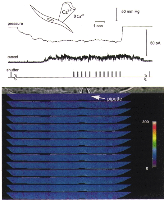
Figure 4. The focal increase in [Ca2+]i is nearly abolished when stretch-activated channels open with the inward driving force on Ca2+ greatly reduced.

When the patch pipette potential is changed to −120 mV so that the patch membrane potential is 120 mV more positive than the resting potential, the stretch-activated channel currents are outward and the driving force for Ca2+ influx is greatly reduced. There is almost no change in [Ca2+]i under these conditions, indicating that an influx of Ca2+ is required for the observed focal increase at negative patch potentials. When the membrane potential was then returned to 100 mV more negative than the resting potential, the focal elevation in [Ca2+]i was again seen with the opening of stretch-activated channels, as shown in Fig. 3B. The outward currents would cause the whole-cell membrane potential to become more negative, decreasing the outward driving force for the unitary currents, so the unitary current amplitude declines with time. Because at positive patch potentials the stretch-activated channel openings are of longer duration (Kirber et al. 1988), this decline in amplitude is reflected in the unitary current while the channel is still open, giving rise to the decay in the current (see Barry & Lynch, 1991). In addition, there may be a contribution to the current record by stretch-activated K+ channels, which can continue to be active after membrane stretch has ceased (Ordway et al. 1995), as can, at times, the stretch-activated cation channels. The experimental conditions were the same as for Fig. 3. The colour bar provides a linear scale for [Ca2+]i from 0 to 300 nM.
