
Figure 7. The hilus is necessary for the post-FPI enhancement of granule cell responses.
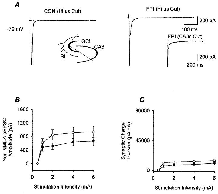
A, means of representative traces from granule cells in slices from control (CON) and FPI animals with the hilus largely removed (as illustrated in the schematic drawing in the inset in the left panel; GCL, granule cell layer; St, stimulating electrode; see Methods), illustrate that, after the removal of the hilus, the late inward current shown in Fig. 4 from FPI animals could not be observed. Recordings were obtained in the presence of bicuculline (20 μM) and APV (20 μM). Note also that the second peak seen in granule cells from ‘uncut’ (i.e. hilus intact) slices from control animals (Fig. 4A) is reduced in the ‘hilus cut’ slices from control animals. The inset in the right panel shows that removal of the CA3c alone (i.e. cutting along the two lines which connect the tip of the CA3c with the tips of the granule cell layer, without removal of the hilus) did not eliminate the late inward current in slices from FPI animals (compare with Figs 4A and 7A); however, there appeared to be a reduction, albeit a non-significant one, in the late EPSC at all stimulation intensities (e.g. at 4 mA stimulation, the charge transfer in granule cells from intact slices from FPI animals was 69 395 ± 13 780 pA ms, as shown in Fig. 4C, versus 39 179 ± 16 183 pA ms in granule cells from ‘CA3c cut’ FPI slices; by comparison, the charge transfer in granule cells from hilus cut FPI slices at the same stimulation intensity was 13 522 ± 1765 pA ms, as shown in Fig. 7C). B, summary data showing the lack of a difference between the amplitudes of the monosynaptic EPSCs in granule cells from slices with the hilus removed from control (•, n = 9) and FPI (○, n = 6) animals. C, the post-FPI enhancement of the synaptic charge transfer shown in Fig. 4E was also eliminated in slices with the hilus largely removed.