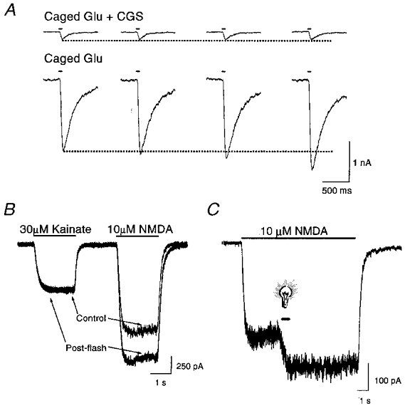
Figure 1. NMDA-activated responses are potentiated by light.

A, whole-cell currents (membrane potential: -60 mV in this and all subsequent figures) in a rat cortical neurone in vitro in response to photolysis (50 ms, > 280 nm) of caged glutamate (Glu, 30 μM) in the presence and absence of 30 μM CGS-19755 (CGS). Sample traces represent consecutive stimuli, 60 s apart, in a single neurone. Similar effects were seen in at least 4 additional cells. B, whole-cell currents elicited by 30 μM kainate and 10 μM NMDA in a cortical neurone before and shortly after (≈5 s) a brief light stimulus to the cell in the absence of caged compounds. Traces are representative of a total of 4 cells. C, whole-cell response of a neurone exposed to light (600 ms, > 280 nm; light bulb symbol) during the application of 10 μM NMDA. Similar effects were seen in 4 additional cells.
