
Figure 6. Difference in excitatory responses produced by transmural nerve stimulation between the parenchyma and sac of the rat penile bulb.
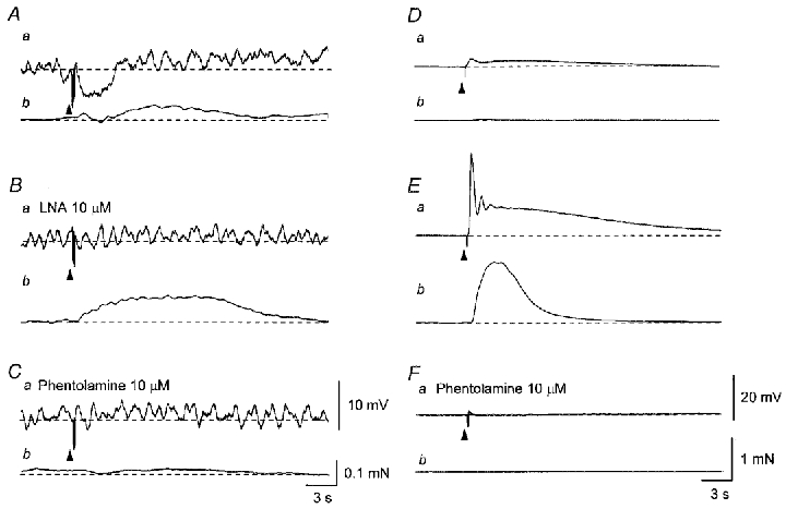
In a parenchyma preparation, five impulses delivered at 20 Hz initiated an IJP (Aa) and a contraction which was interrupted by a relaxation (Ab). In the presence of LNA (10 μM) five impulses delivered at 20 Hz triggered an enhanced contraction without detectable membrane potential changes (Ba and b). Subsequent addition of phentolamine (10 μM) abolished the contraction (Ca and b). In the sac, a single impulse initiated an initial depolarisation which was followed by a slow depolarisation (Da) and also produced a tiny contraction (Db). Three impulses delivered at 20 Hz initiated an initial depolarisation which trigged an action potential and a slow depolarisation (Ea). The action potential caused a large contraction (Eb). Phentolamine (10 μM) abolished both the depolarisation and contraction (Fa and b). Traces in A–C and D–F were recorded from two different preparations. Resting membrane potentials were -36 mV in A–C and -56 mV in D–F. Scale bars in C also refer to corresponding traces in A and B. Scale bars in F also refer to corresponding traces in D and E.