
Figure 7. Effects of nifedipine on responses produced by transmural nerve stimulation in the sac of the rat penile bulb.
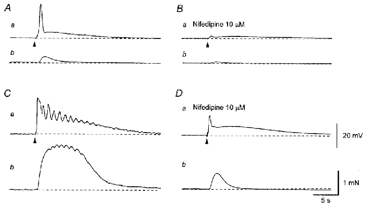
A single impulse evoked an initial depolarisation which triggered an action potential and a slow depolarisation (Aa). The action potential caused a small contraction (Ab). Nifedipine (10 μM) abolished an action potential leaving an EJP, which consisted of two components (Ba). Nifedipine also inhibited the nerve-evoked contraction (Bb). Three impulses delivered at 20 Hz initiated a larger depolarisation and triggered multiple action potentials (Ca) and an associated larger, oscillatory contraction (Cb). Nifedipine (10 μM) abolished action potentials leaving two components of the EJP (Da). Nifedipine also inhibited the nerve-evoked contraction (Db). All traces were recorded from the same cell. Resting membrane potential was -52 mV. Scale bars in D refer to corresponding traces in A–C.