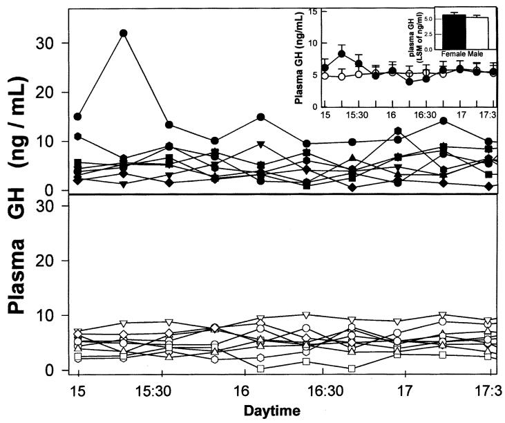
Figure 3. Ultradian GH secretory patterns in dogs. Main panels: Individual ultradian plasma GH profiles for females (lower panel) and males (upper panel). Large inset: Average ultradian GH profiles in male (●) and female (○) dogs. Small inset: Comparison between integrated (pooled data for each gender) plasma GH levels in male and female dogs. Bars over columns represent values for standard error of mean.

An official website of the United States government
Here's how you know
Official websites use .gov
A
.gov website belongs to an official
government organization in the United States.
Secure .gov websites use HTTPS
A lock (
) or https:// means you've safely
connected to the .gov website. Share sensitive
information only on official, secure websites.
