
Figure 10. Immunofluorescence localization of P2X2 subunits and neuronal/neuroendocrine markers in petrosal ganglia and carotid body in situ and dispersed cells in vitro.
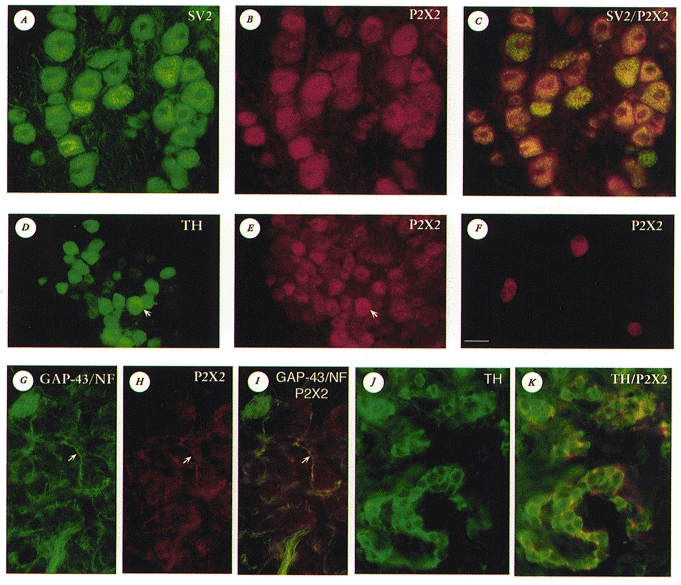
A-C, the same microscopic field from a tissue section of rat petrosal ganglion after immunostaining for SV2 (A; FITC fluorescence) and P2X2 subunits (B; Cy3 fluorescence); in C, dual exposure shows coincident staining for SV2 and P2X2 (yellow-orange fluorescence) in the majority of neurones. D and E, the same microscopic field from a tissue section of rat petrosal ganglion (near exit of the glossopharyngeal nerve) after immunostaining for TH (D; FITC fluorescence) and P2X2 subunits (E; Cy3 fluorescence). Note that many TH-positive neurones, the majority of which innervate carotid body type 1 cells, showed strong P2X2 immunoreactivity, e.g. neurone indicated by arrow in D and E; also, several P2X2-positive neurones were TH-negative (e.g. neurones in top right corner of E). F, petrosal neurones from a 4-day-old culture stained positive for P2X2 immunoreactivity; background cells, not visible in this dark field micrograph, are unstained. G–I, the same microscopic field from a tissue section of rat carotid body after immunostaining for NF/GAP-43 (G; FITC fluorescence) and P2X2 subunits (H; Cy3 fluorescence); note prominent nerve fibre staining for both NF/GAP-43 and P2X2 (e.g. arrows in G–I). Coincident staining is revealed in I by the occurrence of yellow fluorescence. J and K, the same microscopic field from a tissue section of rat carotid body after immunostaining for TH (J; Alexa 488 fluorescence) and P2X2 subunits (Cy3 fluorescence); dual exposure reveals both Alexa 488 and Cy3 fluorescence (K). In K, strong P2X2-positive nerve terminals (red) are intimately associated with TH-positive type 1 cells (green). Note type 1 cells are negative for P2X2 in K. The calibration bar shown in F (lower left) represents: 25 μm in A-C, F, J and K; 30 μm in D, E and G–I.