
Figure 4. Baf reduces evoked EPSC and mEPSCs in autaptic cultures.
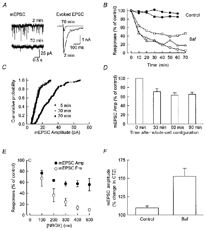
A, sample traces of mEPSCs (left) and evoked EPSCs (right) at 2 and 70 min after whole-cell configuration was established in an autaptic neuron. Baf was present in the recording pipette. Note the dramatic reduction in the amplitude and frequency of mEPSCs and a large reduction in the size of the evoked EPSC. B, time course of the change in mEPSCs and evoked EPSC from one representative cell. The abscissa indicates the time after whole-cell configuration was obtained. Note that the reduction of mEPSCs amplitude (□) plateaued around 30–40 min, while the reduction in mEPSC frequency (▵) and evoked EPSC amplitude (○) continued. The amplitudes of evoked EPSCs (•) and mEPSC s (▪) from a control cell are also shown on the same graph for comparison. The dramatic reduction in evoked EPSCs and mEPSC amplitude and frequency are not caused by rundown of the synaptic responses as a result of long recording. For control cells, the same concentration of DMSO was added to the internal solution. C, cumulative probability distribution of mEPSCs. A clear leftward shift is seen as Baf diffuses into the neuron, while the distribution of mEPSCs at 30 and 70 min are similar to each other. D, the effect of Baf appears to be independent of neuronal activity. The time course of reduction in mEPSC amplitude is shown for four autaptic cells, which were held at -70 mV during the entire course of recording. The time course of the reduction of mEPSC amplitude is similar to that shown in B, which suggests that neuronal activity (action potentials or activity-dependent turnover of synaptic vesicles) is not required for the effect of Baf (71 ± 6 % (30 min), 63 ± 5 % (60 min), and 65 ± 5 % (90 min) of control values). E, the effect of increasing the concentration of NBQX on mEPSC amplitude and frequency. At low concentrations, NBQX causes a reduction in both amplitude and frequency. However, the reduction in amplitude plateaus while the decrease in frequency continues with higher concentrations of NBQX. These results point out the limitation in the analysis of mEPSCs in terms of interpreting the changes in both amplitude and frequency. F, the increase of mEPSC amplitude by CTZ is larger in neurons treated with Baf. mEPSCs were collected in the normal solution and in the presence of 200 μM CTZ, for both control and Baf-treated cells. The relative change of mEPSC was calculated for each cell. The increase in mEPSC amplitude caused by CTZ is larger for Baf-treated cells (mean = 153 ± 11 %, n = 7) than control cells (mean = 110 ± 3 %, n = 10). This result indicates that Baf causes a significant reduction in the concentration of glutamate in the vesicles. Confluent cultures were used for experiments shown in E and F.