
Figure 1. Dendritic arbor (red) and axonal projection (blue) of a layer 4 spiny stellate cell (left) and a layer 5 pyramidal cell of a postnatal day 14 (P14) rat.
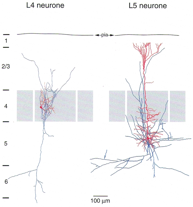
The dendrites of the spiny stellate cell are confined to layer 4 (grey). In rodent barrel cortex dendrites remain exclusively within the cortical barrel in which the soma is located and often show a highly asymmetrical orientation towards the centre of the barrel. The spiny stellate axon collaterals are predominantly vertically oriented. The axonal arbor spans the entire cortex, projecting to layer 1 and to the white matter. Most boutons are on axon collaterals within layer 4 and layer 2/3 (Lübke et al. 2000). The prominent apical dendrite of layer 5 pyramidal cells extends into layer 1 in a terminal tuft. The basal dendrites extend within layers 5 and 6. The axon is long (1-4 mm) and collaterals are mainly horizontally oriented. Fewer vertical collaterals project to layer 1 where they may form synapses with the terminal tufts of other pyramidal neurones.