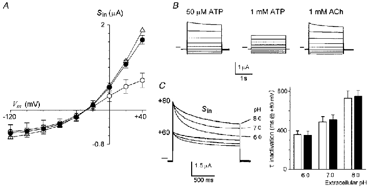
Figure 3. Effects of extracellular ATP and pH on Sin.

A, current-voltage relations of Sin elicited by 50 μM ATP (•), 1 mM ATP (○), or 1 mM ACh (▵). Data are from 5–13 follicles (2-3 frogs) in each condition held at -60 mV. B, superimposed current traces representative of the Sin elicited by the agonists at membrane potentials from +40 to -120 mV. Horizontal lines indicate zero current. C, superimposed current traces obtained at the peak of Sin elicited by 50 μM ACh at the membrane potentials stepped to +80 and +60 mV from a holding potential of -60 mV. At each potential the set of current traces corresponds with Sin generated in extracellular medium buffered to pH 8, 7, or 6. The inactivation kinetics at +80 mV were fitted to simple exponentials and the mean of the time constants (τ) in each value of extracellular pH were plotted in the bar graph. The open bars are the mean τ inactivation for the ICl,swell generated by HR80 (3 follicles, 2 frogs), while the filled bars are those for the Sin elicited by 50 μM ACh in HR90 (7 follicles, 2 frogs) in follicles from the same frogs.
