
Figure 3. Measurements of cAMP concentration.
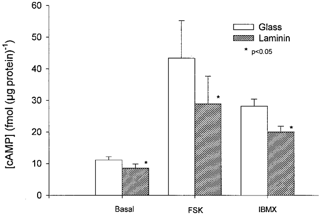
cAMP concentration in cells plated on glass (open bars) or laminin (hatched bars) under basal conditions, stimulated by exposure to 1 μm forskolin (FSK) or by exposure to 100 μm IBMX. Cells plated on laminin exhibited significantly smaller basal, forskolin- and IBMX-stimulated cAMP levels than cells plated on glass.