
Figure 6. Effects of 1 μm ACh on basal ICa,L recorded from atrial cells plated on glass (A), laminin (B) or laminin plus 20 μm cytochalasin D (C).
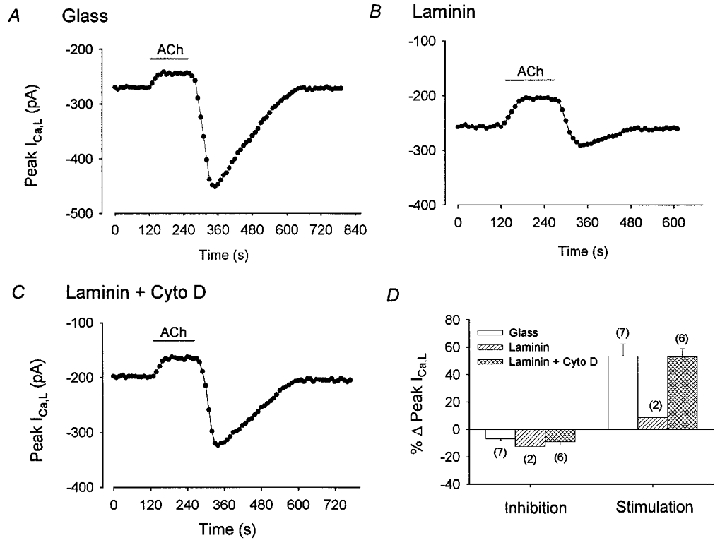
A, B and C, consecutive measurements of peak ICa,L. On glass, ACh induced a typical inhibition and rebound stimulation of ICa,L (A). On laminin, ACh exposure inhibited ICa,L and ACh withdrawal elicited an attenuated stimulation of ICa,L (B). Addition of cytochalasin D to the external solution of cells plated on laminin, restored the rebound stimulation of ICa,L elicited by ACh withdrawal. D, bar graph summarizing ACh-induced inhibition and rebound stimulation of ICa,L in the 3 groups. The numbers in parentheses indicate the number of cells studied in each group.